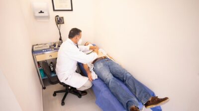
A diabetic eye exam is a comprehensive dilated retinal evaluation that screens for diabetic retinopathy, macular edema, cataracts, and glaucoma. Most exams take 30-60 minutes and include visual acuity, dilation, slit-lamp exam, and digital retinal imaging. The American Diabetes Association recommends a baseline exam at type 2 diabetes diagnosis and annually thereafter — Medicare and most commercial insurers cover it.
Why the Exam Matters
Diabetic retinopathy is the leading cause of vision loss in adults aged 20-74. The danger is that early retinopathy has no symptoms — by the time you notice blurry vision or floaters, significant damage may already be present. Annual exams catch the disease at a stage when laser treatment, anti-VEGF injections, or tighter glucose control can preserve sight.
What the Exam Includes
| Test | What It Measures | Time |
|---|---|---|
| Visual acuity | Sharpness of vision (Snellen chart) | 2-3 min |
| Intraocular pressure (tonometry) | Screens for glaucoma | 2 min |
| Slit-lamp exam | Front of the eye: cornea, iris, lens (cataracts) | 5 min |
| Dilation | Drops widen the pupil for retina view | 15-20 min wait |
| Dilated fundus exam | Direct retina visualization with ophthalmoscope | 5-10 min |
| Fundus photography | Wide-field digital photo of retina | 5 min |
| Optical coherence tomography (OCT) | Cross-section image of retinal layers; detects macular edema | 5-10 min |
| Fluorescein angiography (when indicated) | IV dye highlights leaky blood vessels | 20-30 min |
Step-by-Step: What Happens at Your Appointment
- Check-in and history. The technician asks about diabetes duration, A1C, medications, and any vision changes.
- Visual acuity. You read letters from a chart with each eye covered separately.
- Tonometry. A puff of air or a small probe measures eye pressure.
- Dilation. Drops are placed in each eye; the technician asks you to wait 15-20 minutes for the pupils to widen.
- Slit-lamp and fundus exam. The doctor examines the front of the eye, then the dilated retina with a bright light and lens.
- Imaging. Fundus photographs and OCT scans provide a baseline that future exams can compare against.
- Discussion. The doctor reviews findings, shows you the images, and recommends a follow-up interval.
Frequency of Exams
| Situation | Recommended Interval |
|---|---|
| Type 2 diabetes, newly diagnosed | At diagnosis, then every 1-2 years if no retinopathy |
| Type 1 diabetes, post-puberty | 5 years after diagnosis, then annually |
| Mild non-proliferative retinopathy | Every 6-12 months |
| Moderate to severe retinopathy | Every 3-6 months |
| Proliferative retinopathy or macular edema | Every 1-3 months, often with treatment |
| Pregnancy with pre-existing diabetes | Before conception, in the first trimester, then as advised |
| Prediabetes | Routine eye exam every 1-2 years; specific diabetic exam not required |
What Doctors Look For
- Microaneurysms — earliest sign of retinopathy
- Dot and blot hemorrhages — small areas of bleeding in the retina
- Hard exudates — yellow lipid deposits
- Cotton-wool spots — areas of nerve fiber damage
- Macular edema — fluid swelling in the central retina (most common cause of vision loss)
- Neovascularization — abnormal new blood vessels (proliferative retinopathy)
- Cataracts and elevated eye pressure — both more common in diabetes
Cost and Insurance Coverage
| Coverage | Detail |
|---|---|
| Medicare Part B | One annual exam covered; 20% coinsurance after Part B deductible |
| Medicare Advantage | Often includes routine vision and waives the 20% copay |
| Commercial medical insurance | Most cover annual diabetic eye exam under medical (not vision) benefit |
| Vision insurance (separate) | Covers refraction and glasses; typically not the dilated medical exam |
| Self-pay | $100-$250 for the exam; OCT may add $50-$150 |
Tips for the Day Of
- Bring a list of medications, recent A1C, and the name of your prescribing physician.
- Bring sunglasses — dilated pupils stay sensitive to light for 4-6 hours.
- Arrange a ride if you have never been dilated before.
- Avoid scheduling near-vision tasks (reading, fine close work) for the rest of the day.
- If you wear contact lenses, bring your case and solution; you may be asked to remove them.
When to See an Eye Doctor Sooner
Call your eye doctor — do not wait for the annual visit — if you notice:
- Sudden vision loss in one or both eyes
- New floaters or flashing lights
- A dark spot or “curtain” in your visual field
- Sudden blurring that does not clear with blinking
- Pain, redness, or persistent eye discomfort
Vision change can also be an early symptom of prediabetes or rising blood sugar. See our companion article on diabetes eye symptoms for more on what these changes can mean.
According to the American Academy of Ophthalmology, early detection and treatment can reduce the risk of severe vision loss from diabetic retinopathy by more than 90%.
The Bottom Line
A diabetic eye exam is a comprehensive dilated retinal evaluation — far more thorough than a routine vision check. Get one at diagnosis if you have type 2 diabetes, 5 years after diagnosis if you have type 1, and annually thereafter. Medicare and most commercial plans cover it. Catching retinopathy early — before symptoms appear — is the single most effective way to protect long-term vision.