Glucose is a six-carbon sugar with the molecular formula C6H12O6. Its chemical picture shows two main forms: an open-chain aldehyde and a six-membered ring (pyranose) formed when carbon 1 connects to the oxygen on carbon 5. In solution — the form that matters in blood, urine, and body fluids — more than 99 percent of glucose exists as the ring. The biologically active form is D-glucose; its mirror image (L-glucose) is synthetic and not metabolized.
The Open-Chain Structure
The open chain of D-glucose is drawn with six carbons in a vertical line:
- Carbon 1 (C1) has an aldehyde group (-CHO)
- Carbons 2, 3, 4, 5 each have a hydroxyl group (-OH)
- Carbon 6 (C6) has a hydroxyl group plus the terminal hydrogens
This open-chain form accounts for less than 1 percent of glucose in water. It is mostly a transient state.
The Ring Structure: Pyranose
When glucose dissolves in water, the aldehyde on carbon 1 reacts with the hydroxyl on carbon 5, forming a six-membered ring that contains one oxygen atom. This ring form is called a pyranose. Most chemistry diagrams show it as a hexagon lying on its side (the Haworth projection) or as a chair conformation (more realistic 3D shape). Key features:
- Five carbons and one oxygen form the ring
- Hydroxyl groups stick up or down from each ring carbon
- The carbon outside the ring (C6) carries a CH2OH group
Alpha and Beta Anomers
When the ring forms, carbon 1 can end up with its new hydroxyl either below (alpha) or above (beta) the plane of the ring. These are called anomers and interconvert slowly in solution:
- Alpha-D-glucose: ~36 percent of glucose in solution
- Beta-D-glucose: ~64 percent of glucose in solution
- Open chain: less than 1 percent
The two anomers mix in what chemists call mutarotation. Both are used by cells, though some enzymes prefer one over the other.
D-Glucose vs L-Glucose
| Feature | D-Glucose | L-Glucose |
|---|---|---|
| Occurrence | Plants, animals, fungi; all life | Synthetic only |
| Metabolized by humans | Yes — primary fuel | No — not recognized by most enzymes |
| Structure | Mirror image of L-glucose | Mirror image of D-glucose |
| Taste | Sweet | Sweet (to humans; not used for energy) |
| Source | Fruit, honey, cornstarch, blood | Laboratory synthesis |
When medical references say “glucose,” they mean D-glucose unless otherwise specified.
Where Glucose Is Found
- Blood: the form measured as “blood sugar,” “plasma glucose,” and “fasting glucose” — all D-glucose
- Cells: the fuel for glycolysis and the citric acid cycle, producing ATP
- Liver and muscle: stored as glycogen, a long chain of glucose units
- Food: starch (a plant glycogen equivalent) breaks down to glucose; sucrose (table sugar) is glucose bonded to fructose; lactose (milk sugar) is glucose bonded to galactose
- Urine: normally trace amounts; significant glucose in urine (glycosuria) indicates high blood glucose spilling past kidney reabsorption
- Interstitial fluid: the form CGMs measure
Why the Shape Matters
Enzyme Recognition
Enzymes like hexokinase, glucokinase, and glucose-6-phosphatase recognize D-glucose specifically. The ring shape and the arrangement of hydroxyl groups provide the exact 3D surface these enzymes need to bind and catalyze. L-glucose simply does not fit.
Sweet Taste
Sweet taste receptors on the tongue recognize specific 3D features of sugars. Both D- and L-glucose trigger these receptors, which is why L-glucose tastes sweet even though the body cannot use it.
Drug Design
The shape of glucose is also why SGLT2 inhibitors (empagliflozin, dapagliflozin, canagliflozin) work. These drugs mimic glucose’s 3D structure enough to bind to the SGLT2 transporter in the kidney tubule, blocking glucose reabsorption and causing it to be excreted in urine. The drug design depends on understanding the glucose shape precisely.
Glucose Versus Other Sugars
| Sugar | Structure | Digestion Path |
|---|---|---|
| Glucose | Six-carbon monosaccharide | Absorbed directly |
| Fructose | Six-carbon monosaccharide (ring is five-membered furanose) | Absorbed, processed by liver; different metabolic effects |
| Galactose | Six-carbon monosaccharide | Converted to glucose in liver |
| Sucrose | Glucose + fructose disaccharide | Split into glucose and fructose in gut |
| Lactose | Glucose + galactose disaccharide | Split into glucose and galactose by lactase |
| Starch | Long chain of glucose units | Broken down by amylase into glucose |
| Glycogen | Branched chain of glucose (animal storage form) | Broken down to glucose on demand |
| Cellulose | Long chain of glucose with different bonds | Not digested by humans (dietary fiber) |
How the Body Measures “Glucose”
- Fasting plasma glucose: venous draw after 8 to 12 hours of fasting
- Random plasma glucose: venous draw at any time
- OGTT: timed series of draws before and after a 75 g or 100 g glucose drink
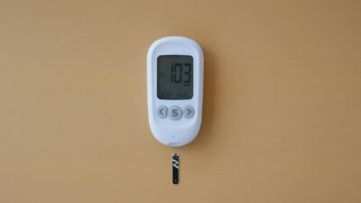
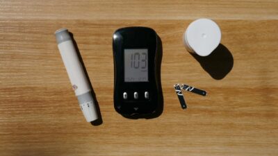
- Fingerstick glucose: capillary drop read on a home meter
- Continuous glucose monitor (CGM): interstitial fluid glucose every 1 to 5 minutes via subcutaneous sensor
- A1C: not a glucose measurement directly — it measures glycated hemoglobin as a proxy for average glucose over 2 to 3 months
See our glucose equation reference for how these measurements relate mathematically.
Glucose in Disease
Diabetes
In diabetes, blood glucose rises above normal because of insulin deficiency (type 1) or insulin resistance (type 2). The diagnostic cutoffs are based on D-glucose concentration:
- Normal fasting: under 100 mg/dL
- Prediabetes: 100 to 125 mg/dL
- Diabetes: ≥ 126 mg/dL (repeated)
Hypoglycemia
Low blood glucose (under 70 mg/dL) causes adrenaline-driven warning symptoms and, if severe, neurologic dysfunction. See our guide on treating low blood sugar.
Hyperglycemia
Chronic elevations damage small and large blood vessels, driving the long-term complications of diabetes — retinopathy, nephropathy, neuropathy, and cardiovascular disease.
Visualizing Glucose in Your Daily Life
- Every time you eat carbs, starches break down to glucose in your gut.
- Glucose enters the bloodstream and triggers insulin release from the pancreas.
- Insulin signals muscle and fat cells to absorb glucose and store or burn it.
- The liver stores excess glucose as glycogen for later release between meals.
- The brain uses roughly 120 g of glucose per day — about 60 percent of the body’s baseline glucose use.
- CGM sensors and lab tests both measure D-glucose concentration at different sample sites.
Related Reading
See our guides on the glucose equation, A1C levels, and random glucose testing for how glucose measurement fits into clinical care.
The Bottom Line
The “glucose picture” is a six-carbon ring-shaped sugar (D-glucose, C6H12O6) that exists mostly as a pyranose ring in water. Its specific shape is why enzymes recognize it, why the body uses it as primary fuel, and why medications targeting glucose transport can work so precisely. Every lab test, home meter, and CGM is measuring the same underlying D-glucose molecule — just in different body fluids and at different time scales.